Glaucoma or blue water is a disease that may lead to optic nerve defect and consequently, blindness. The disease has no symptoms at the beginning; however, it can reduce vision and eventually blindness over a few years. Early treatment may prevent its progression and reduction in patient's vision.
Optic Nerve:
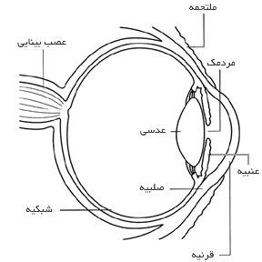
Optic nerve consists of over one million neurons come together in a series of strands. This nerve connects retina (a thin, light sensitive screen back of the eye) to the brain (Fig. 1). The healthy optic nerve is essential for a good vision.

Fig. 1
The Impact of Glaucoma on the Optic Nerve:
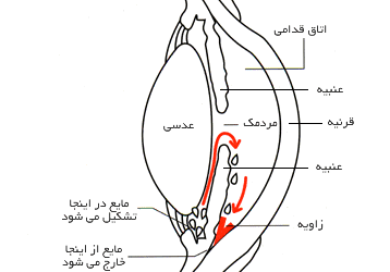
In many individuals, increased intraocular pressure results in glaucoma. There is a space called “anterior chamber” in front of the eye. A transparent fluid always enters this space and gets out of it, and it is responsible for feeding the adjacent tissues. The site where the fluid enters after getting out of the anterior chamber is called the “angle” that is the intersection of cornea and iris (Fig. 2). When the fluid reaches the angle, a spongy network like a drainage system gets out of the eye.

Fig. 2
Open-angle glaucoma is referred to by this term since the angle from which the fluid gets out is “open”. However, for unclear reasons, the flowing speed of the fluid through the drainage network is low. The slow flow of the fluid leads to its accumulation and consequently the increased intraocular pressure; this increase will damage the visual nerve and reduced vision unless the intraocular pressure is lowered.
Who are at risk?
• Individuals over 40 years old
• Individuals with a history of this disease in their family
Symptoms of Glaucoma Disease:
Open-angle glaucoma is symptom less at first. Vision is normal and there is no pain. As the disease progresses, the patient notices that although he well sees the objects in front of him, he could not see well the objects next to him that he should look at them from the corner of his eye.
If left untreated, the patient with glaucoma may suddenly realize that he has no “side vision”, just like looking around from inside a pipe. Progress of the disease may cause the remaining vision to be disappeared even in the center and consequently, the patient becomes blind (Figs. 3 and 4).
 |  |
| Normal Vision | Vision of a Patient with Glaucoma |
Diagnosis of Glaucoma:
Many people think that they have glaucoma only if their intraocular pressure is high; however, this is not always true. High intraocular pressure increases the risk of glaucoma. Nevertheless, high intraocular pressure does not necessarily mean glaucoma.
Having or not having glaucoma due to the high intraocular pressure depends on the rate of endurance of optic nerve against the high intraocular pressure; and this rate is different in different people. Although the normal intraocular pressure is usually between 12 and 21 mmHg, an individual may have glaucoma even with this pressure, indicating that eye examination is very significant.
To diagnose glaucoma, the ophthalmologist should perform the following diagnostic measures:
• Visual Acuity: In this test, performed by means of visual charts, the patient's vision is determined at different intervals.
• Visual field: In this test, the patient's corner (peripheral) vision is measured. Considering that loss of peripheral vision is one of the glaucoma symptoms, this test contributes to the diagnosis of the disease.
• Dilated pupil: In this test, the patient’s eye is dilated using drops, so that the ophthalmologist finds a better vision to examine the visual nerve. After examination, the near vision may be blurred for a few hours.
• Tonometry: In this test, the intraocular pressure is measured.
Glaucoma Treatment:
Although glaucoma has no definite treatment, it is controlled by the current treatment, confirming the importance of early diagnosis and treatment. Most ophthalmologists treat the newly diagnosed glaucoma with medication; nevertheless, new researches have revealed that laser surgery is a safe and effective alternative.
Treatment procedures for glaucoma include:
• Medical Treatment: medical treatment is the most common type of early treatment for glaucoma. Glaucoma medicines are prescribed as eye drops and tablets. These medications decrease the intraocular pressure in two ways. Some reduce the fluid production in the eye, and some help drain more fluid from it. Anti-glaucoma drugs may be prescribed up to several times a day. Most patients show no side effects; however, some of them may cause headaches or complications on other organs in the body. Drops may lead to irritation and reddening of eyes. Anti-glaucoma drugs should be used as long as they help control intraocular pressure. Since glaucoma is not usually associated with symptoms, sometimes patients stop using or forget their drug.
• Laser Surgery: Laser surgery contributes to drainage of fluid from the inside of the eye. Although this method may be used at any time, it is usually employed after testing medication treatment. In many cases, the patient should take drugs even after laser surgery.
• Common Surgical Procedures: the goal in glaucoma surgery is to create a new outlet for the fluid in the eye. Although an ophthalmologist may decide to perform surgery at any time, he usually does it after the failure of medication and laser surgery. Surgery is performed in the clinic or hospital. Before surgery, relaxation drugs are given to the patient, and then the eye is benumbed by injecting anesthetic substances around it.
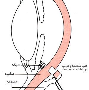
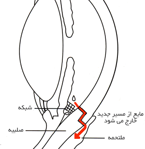
The surgeon eliminates a small piece of the white tissue of the eye (sclera), creating a small canal for the passage of fluid flow inside the eye. Then the removed white piece of the eye is covered with a thin, clear layer of conjunctiva. Liquid gets out of the eye from the duct and under the conjunctiva covering it. (Figs. 5 and 6)

Fig. 5
The patient must use antibiotic and anti-inflammatory drops for several weeks after the surgery, to cope with infection and swelling. It should be noted that these drops are different from the ones previously used by patient to treat glaucoma. Moreover, the patient should be visited regularly, in particular during the first few weeks after the surgery.
In some patients, surgery is effective in reducing the intraocular pressure about 80 to 90 percent. However, if the new duct formed during surgery is blocked, the patient may require another surgery. In case that the patient has not already undergone eye surgery (like cataract surgery), glaucoma surgery is associated with the best effect.
It should be considered that although the glaucoma remains the patient remaining vision, it would not improve vision. In fact, the patient's vision may not be well like that of before surgery; however, without surgery, the patient may completely lose his vision in the long term.

Fig. 6
As any other surgery, glaucoma surgery may be associated with side effects including cataracts, corneal problems, inflammation or intraocular infection, as well as the swollen blood vessels in the back of the eye. Of course, there are effective treatments for each of these cases.
Other Types of Glaucoma: Although open-angle glaucoma is the most common type of this disease, glaucoma has other types, too:
• In normal or low-pressure glaucoma, visual nerve degradation and visual restriction unexpectedly occur in people with normal intraocular pressure. The same open-angle glaucoma treatment methods are use in the treatment of these patients.
• In the closed-angle glaucoma, the fluid is located in front of the eye due to the blockage of the angle with the part of the iris (the colored part of the eye); it cannot find a way to the angle and it cannot be removed from the eye. A sudden increase in eye pressure is observed in these patients. The symptoms of this type of glaucoma include severe pain and nausea, and redness of the eye as well as must be immediately treated. If left untreated, the patient may become blind in one or two days. Immediate laser surgery often removes the obstruction and saves the patient from becoming blind.
• In congenital glaucoma, due to a congenital defect at the angle, the child has glaucoma. The affected children usually have obvious symptoms like cloudy eyes, light sensitivity, and severe tearing. Surgical treatment is usually performed, since the anti-glaucoma medications may have unclear impacts on infants, and it is difficult to prescribe these drugs for these patients. If surgery is conducted immediately after diagnosis, the children will have a very high chance of having a good vision.
• Secondary glaucoma may be the complication of other diseases. Sometimes, this type of glaucoma is caused by eye surgery or advanced cataract, eye injuries, some eye tumors, or uveitis (inflammation of the eye). One of the types named pigmentary glaucoma occurs when the pigments are separated from the iris in the form of scales; and by obstructing the outlet network, they prevent the ocular fluid from getting out of the eye. In addition, there is a severe type called neovascular glaucoma that is associated with diabetes. Moreover, corticosteroid drugs used to treat ocular inflammation and other diseases can cause glaucoma in a low percentage of patients.