.jpg)
The cornea is a transparent structure at front of the eye that like a window separates the inside of the eye from the outside. The cornea has two important functions: first, passes light into the eye, and focuses them on the retina, secondly, protects the inner contents of the eye.
In order to allow the light to enter the eye and reach the retina, it must first pass through the cornea. Therefore, the clarity of the cornea plays a key role in vision, and any problem can disrupt the function of this very important member of the visual system. Ophthalmologists of Tehran-Iran Noor Eye Hospital have identified these problems using various diagnostic methods and advised patients to follow the treatment process.
Advanced Cornea Examination and Evaluation Equipment
Today, with the developments of ophthalmology advanced tools, equipment, and techniques have been developed to examine and evaluate the structure and functioning of the cornea. These tools, which are now used in Tehran-Iran Noor Eye Hospital are as following:
1. Topography
In topography a series of concentric light rings is focused on the anterior surface of the patient's cornea and reflected back to a digital camera at the centre of the bowl which the patient is seated facing it and containing the illuminated pattern. Any corneal disorders, such as keratoconus (abnormal curvature of the cornea), corneal scars and also curvature of the cornea can be diagnosed by analyzing the received information and obtained topographical maps. This method is a major measure for people who want to undergo refractive surgeries such as LASIK, LASEK and PRK. It may also be done as post-operative follow-up examination in some people.
2. Pachymetry
Pachymetry is a method in which the thickness of the cornea is measured using ultrasound waves. It also is done before refractive surgery, because in these surgeries a part of the cornea is removed.
3. Pentacam and Orbscan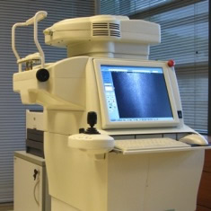
Corneal topography, the cornea's general structure, including surface strength, thickness, and shape of anterior and posterior part of the cornea are determined by pentacam and orbscan. This technique is one of the techniques that can assess posterior part of the cornea.
4. Confoscan
This technique can be used to examine the cornea before surgery, detect early keratoconus, examine the corneal endothelium (the innermost corneal layer), measure the thickness of the corneal layer that is removed in some surgeries, and examine the corneal epithelium (the outermost layer of the cornea ).
5. Slit Lamp Photo
The slit lamp photo has a similar structure to the slit lamp, except that it has a photographic digital camera used by ophthalmologists to take photos of patients' eyes and evaluate them. This device is commonly used to explore the cornea in patients with rare corneal diseases.